

In this Section...
Methods Applied to Orsten Fossils: SEM Work
2. Using scanning electron microscopy (SEM) to study 'Orsten' fossils
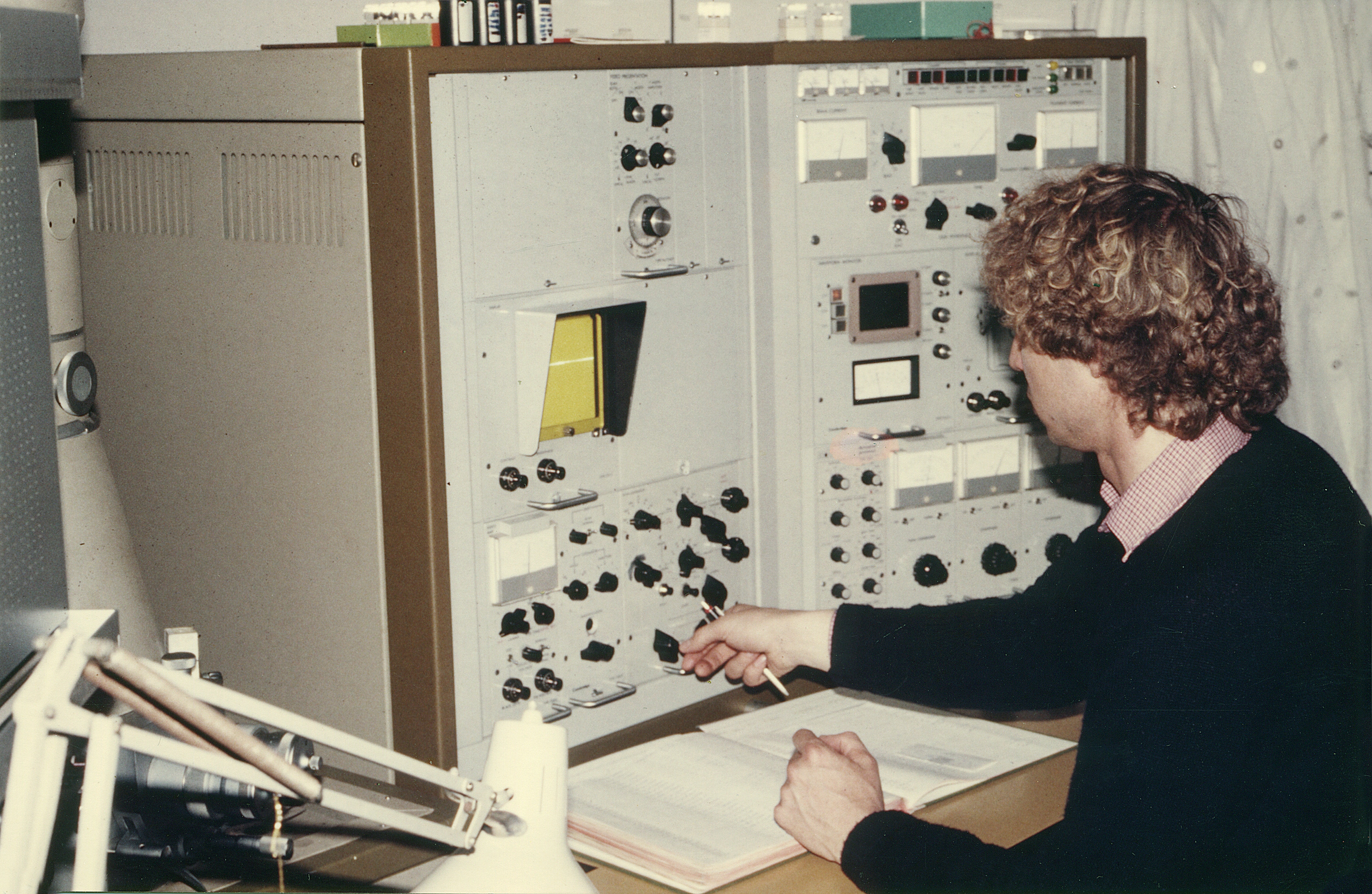
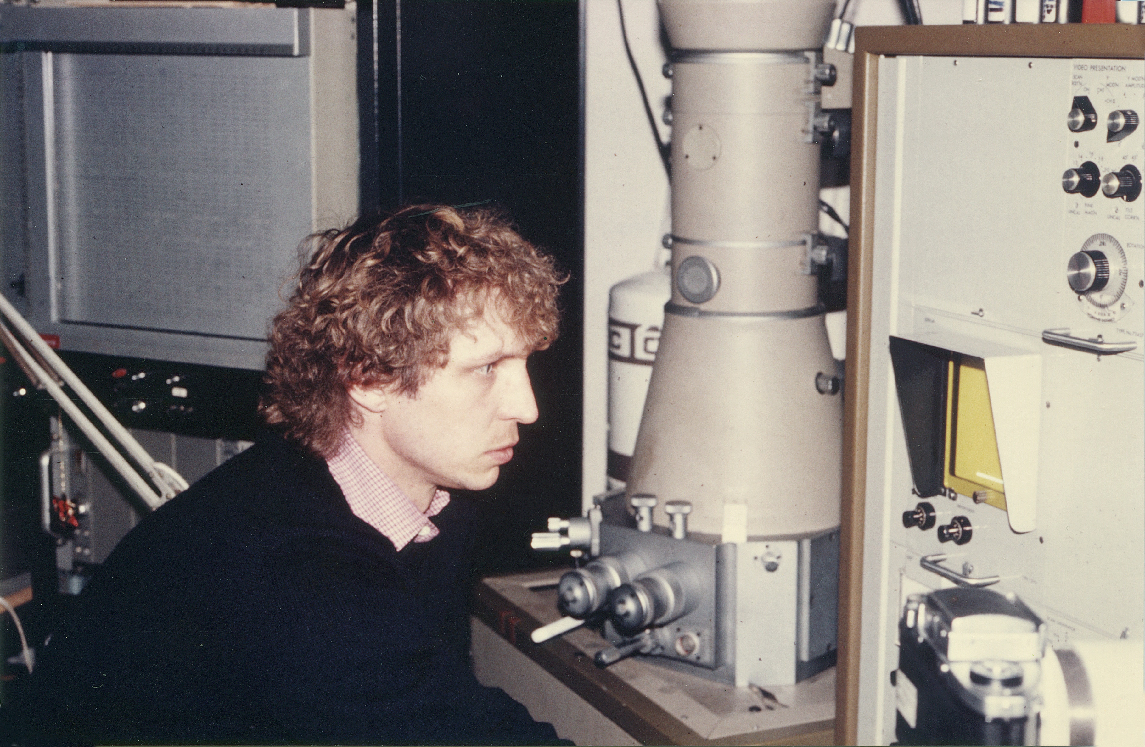
From beginning SEM work became not only our major work tool, but it is our "eye" to view on these extremely tiny critters. As mentioned above, light microscopy yields no results at all. One can see specimens, which are brownish and shiny, but details cannot be seen. Therefore, the only method to visualize such small material, i.e. starting at magnifications at 50 or even 100 times, rarely below, for overviews and from 1,000 times to 10,000 times magnification for details such as the bristles and pores. At this magnification, the granular phosphatic surface contrast and the resolution hinder nice views, but finest pores of 1-2 µm or setulae of diameters below the micron range, i.e. between 0.2 and 0.5 µm have to be documented that way – we used larger lens opening and raised the voltage, when possibly. The normal voltage we used was 10,000. Already the S4 Cambridge SEM (still all valves to be operated manually) allowed to tilt and by this to create series of shots of a specimen at different angles. Combined, one could even yield a, more or less, smoothly running movie. At the beginning the analogous camera allowed pictures of 9 X 9 cm, and each scan took quite a long time (one scan run over an image). We developed all images in a lab, for which Georg Oleschinski was responsible. He also made the final pictures for our tables – all handmade.
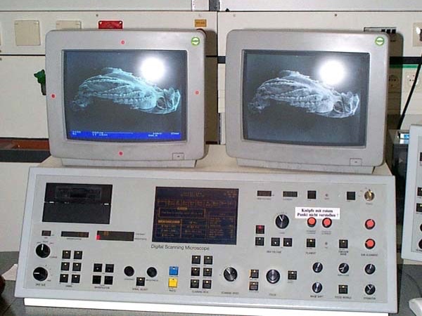
Later in Ulm, we used a SEM at the Center of Electron Microscopy of the university, which had only an extremely old apparatus, but with digital equipment, so we switched to digital images – BUT just 1024 X 1024 pixels. These had to be transferred into a rectangular image to fit the real size. Until Dieter's retirement 2016 we had to stick to that old machine, because the university did not buy any newer apparatus for us. The only good thing: team of the central service unit built a special tilting and rotation unit for us, which permits tilting of 90 degrees. At least that helped much, particularly when re-scanning old specimens of which we have only overviews. Yet this SEM was extremely slow and we could not take as many photographs as we wished. Indeed time ran out to document all the material Klaus had left us: Each year we had to realize that more specimens were destroyed, either single ones cracked and flipped off the stubs, or the entire SEM stub was destroyed – then with 10-20 specimens on it, some specimens figured in publications or even type specimens on it. Big Loss, which affected also various beautiful specimens of Rehbachiella, Skara or Dala.
After Dieter's retirement, he transferred the material, borrowed from the Steinmann Institute, University of Bonn (the real owner, not Klaus) to the University of Lund. His hope is that at some stage more of the material can be documented there. Yet no one is in sight for this job, and also not to continue working up the huge rest remaining in the collection.
© 2020 CORE
